Histology of #Angiogenesis After #Sclerotherapy
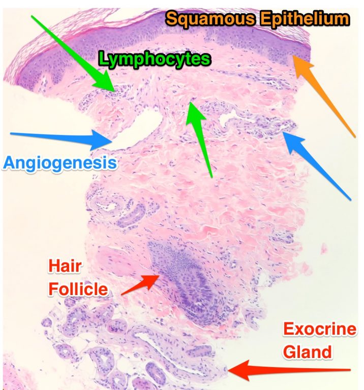
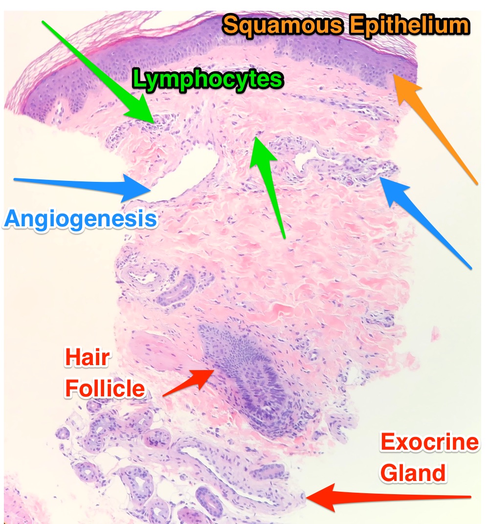
This is a biopsy of a patient that had angiogenesis after sclerotherapy. By studying this image, we can gain a lot of insight into the etiology of angiogenesis. Note that the dilated vessels have hylanized walls. This is secondary to the effects of inflammation on the smooth muscle wall. There are also many leukocytes present surrounding the vessels. These cells are responsible for initiating the inflammatory response. This image is also unique in that it also contains other major components of the dermis ie: exocrine glands and hair follicles.
Learn more about angiogenesis and how to effectively treat and prevent it. Much of the information is based on histological studies which is not available at other ‘Vein Conferences.’